Author:
Kai Knudsen
Reviewer:
Helge BrändströmUpdated:
5 April, 2026
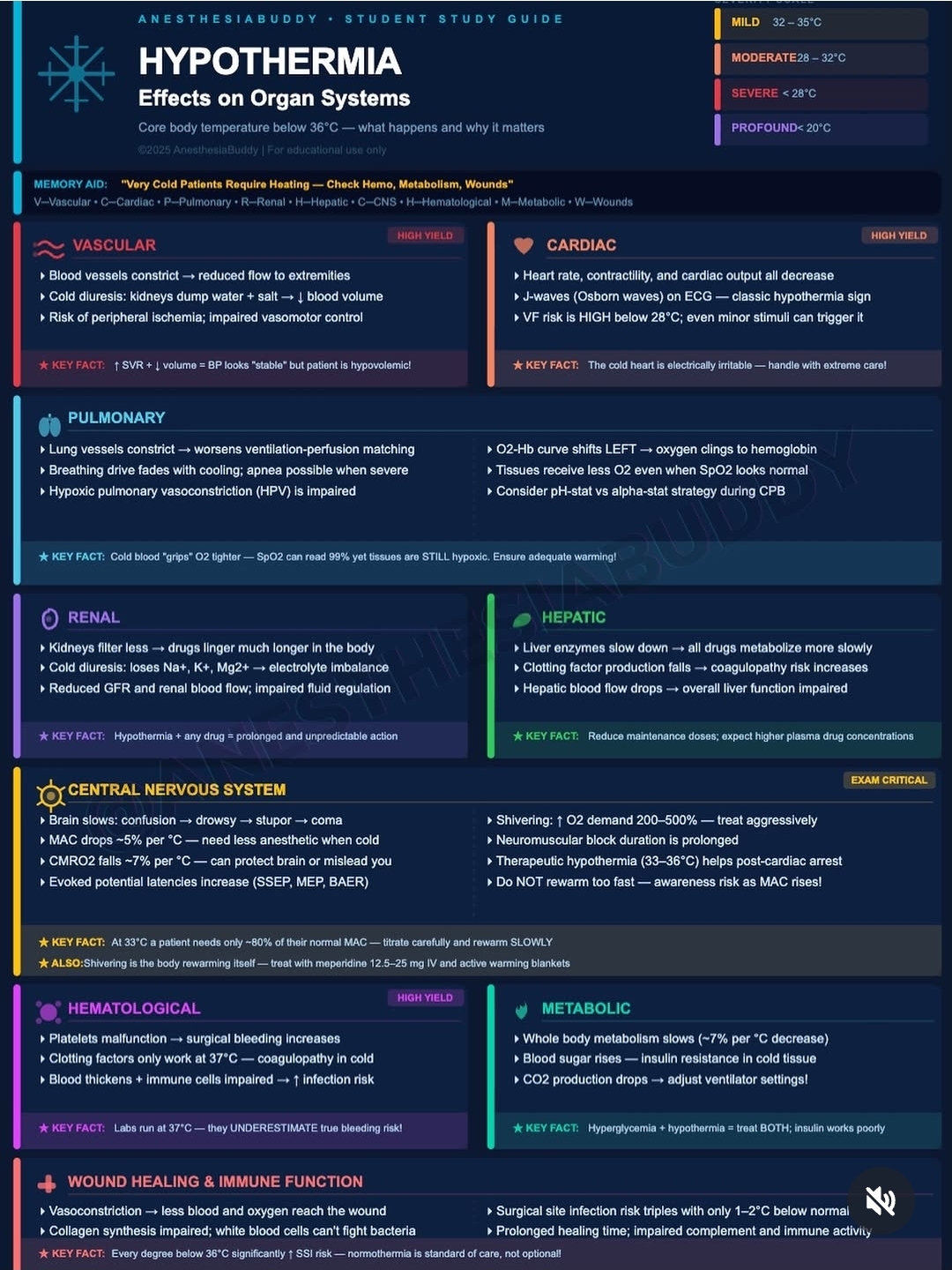
Cooling in an environment with low ambient temperature and/or prolonged exposure where we cannot protect ourselves or produce enough heat to maintain a core temperature of 37°C leads to hypothermia. The medical definition of accidental hypothermia is a body temperature of 35°C or lower.
Hypothermia
Cooling in an environment with low ambient temperature and/or prolonged exposure where we cannot protect ourselves or produce enough heat to maintain a core temperature of 37°C leads to hypothermia. The medical definition of accidental hypothermia is a body temperature of 35°C or lower.
Cold injuries include general cooling (hypothermia) and localized cold injuries. The earliest sign of cooling may be a desire to find warmth. Memory and judgment deteriorate early, speech becomes slurred, and consciousness becomes clouded. Cooling, even at the stage from 37°C down to 35°C, leads to shivering as a way to increase heat production. Shivering is fully autonomously controlled, can increase heat production 2–5 times, and is maximal between 35–33°C. It then decreases with a drop in body temperature, disappearing entirely between 31–30°C.
Early signs include general vasoconstriction in arms and legs, hands, and feet to save central heat, followed by an increased heart rate with additional strain on the heart and an increased respiratory rate. From 35°C and below, the heart gradually cools, leading to a progressive slowing of the pulse and breathing rate. At 34°C, the boundary for hypothermia has clearly been crossed, often with apathy, but sometimes also with psychosis-like symptoms such as hallucinations. Anxiety, restlessness, and increasing effects on consciousness, as well as increasing difficulties with walking, balance, and coordination, are commonly part of the clinical picture and increase as body temperature drops. If the person is not removed from the situation and provided with warmth, body temperature will continue to drop.

At 30°C, most individuals are clearly drowsy, some unconscious. Below a body temperature of 28°C, the heart becomes very sensitive to external stimuli and may enter ventricular fibrillation with rough handling. At 25°C and below, the heart can spontaneously go into asystole or ventricular fibrillation. Hypothermia affects several body functions, including blood clotting ability, and the hypothermic patient becomes increasingly prone to bleeding with a decreasing body temperature. At around 33°C, clotting ability is reduced to about 50% of normal, something evident in both blood tests and clinically. However, a problem is that blood samples are normally analyzed at 37°C, so the results may appear normal while the patient is prone to bleeding. The combination of major trauma, such as simultaneous femur fracture and abdominal bleeding, and hypothermia down to 33°C has been shown to increase the risk of death by up to 40% compared to the corresponding risk in people with normal body temperature. It should be noted, though, that there are large individual differences in how people are affected by cooling.
Initial Management
- Remove wet clothes
- Remove from non-insulated surfaces
- Protect from wind
- Apply warmth
ABCDE in Cold Physiology
- Decreased minute ventilation – EtCO2/PaCO2
- Increased electrical excitability
- Vasoconstriction / Bradycardia
- Coagulopathy
- Altered consciousness
- Modified CPR
- Cold diuresis / Rhabdomyolysis
- Afterdrop / Rescue collapse
A/B – Airway Breathing
- Tachypnea can lead to decreased minute ventilation and apnea.
- Pulmonary edema common after resuscitation from 24°C.
- Increased gradient between PaCO2 and EtCO2
C – Circulation
- Sympathetic activation, tachycardia, hypertension –> bradycardia + peripheral vasoconstriction + hypotension/shock –> 32-28°C.
- Arrhythmias / cardiac arrest
D – Disability
- Altered consciousness, confusion.
- With loss of consciousness / paradoxical undressing 30%.
- At 29°C, dilated, non-reactive pupils.
- At 23°C, absence of corneal reflex (pseudo-rigor mortis / muscle rigidity)
E- Exposure
- Cold diuresis (vasoconstriction-induced hypervolemia + diminished renal tubular responsiveness to ADH)
- Rhabdomyolysis
- Coagulopathy; DIC
- Reduced absorption in GI
- Reduced enzyme activity
- Reduced effect of inotropic drugs
CPR in hypothermia
Refrain from administering medication during Advanced Cardiopulmonary Life Support (A-CPR) if <30°C.
- Between 30°C and 35°C, the interval for medication administration should be doubled, i.e., adrenaline should be given every 8 minutes instead of every 4 minutes. At normothermia (≥35°C), the standard interval in the protocol is used.
- If defibrillation is unsuccessful after 3 attempts, further attempts should be postponed until the core temperature reaches 30°C.
- Between 30°C and 35°C, the interval for medication administration should be doubled, i.e., adrenaline is administered every 8 minutes instead of every 4 minutes.
- At normothermia (≥35°C), the standard interval in the protocol is followed.
Rescue Collapse = Electrical excitability in hypothermia that easily leads to arrhythmias
Afterdrop = Peripheral cold blood reaches central areas during rewarming
ECG in hypothermia
- Bradycardia
- PR prolongation
- Artifacts from shivering
- Various heart blocks
- Atrial fibrillation/flutter
- Widened QRS can lead to prolonged QTc
Asystole (does not rule out a reversible process!)
Osborne J-waves < 32°C (can also arise from sepsis, intracranial pathology, and other causes)
Other causes of J-waves
- Hypercalcemia
- Takotsubo cardiomyopathy
- Left ventricular hypertrophy due to hypertension
- Early repolarization
- Brugada syndrome
- “Le syndrome d’Haïssaguerre” (idiopathic VF – Diagnosis of exclusion in patients who have survived a VF episode without any identified structural or metabolic cause despite extensive diagnostic testing)
Extended Diagnostics for Hypothermia
- Myxedema coma
- Adrenal insufficiency
- Intoxication
- Sepsis
- Pancreatitis
- DKA (Diabetic Ketoacidosis)
Lab Diagnostics for Hypothermia
- Blood status + electrolytes + glucose + ABG
- INR, APT, platelets, fibrinogen
- Blood cultures
- CK, lactate, serum cortisol
- TSH, free T4
The blood gas machine warms samples to 37°C
Expected Abnormalities
- +2% in Hct/°C
- WBC ➡️/↘
- Hemolysis
- Hyperglycemia
- Acid-Base /°C = pK
- Acid-base disturbances are complex, both acidosis/alkalosis can be normal. In warmed samples, the partial pressure of gases increases, and the pH is lower compared to the cold blood in the patient.
- Charles’s Law (also known as the law of volumes) is an experimental gas law that describes how gases tend to expand when heated.
- HCT – Hematocrit increase = due to reduced circulating plasma volume
- WBC – Sequestration
- Hyperglycemia – cold-induced insulin resistance + catecholamine response with gluconeogenesis
- Note! – Warming the sample can mask the true coagulopathy.
Treatment Stage 1-2
Stage 1: 35-32 °C – External heat, movement, warm drink
Stage 1 – Often shivering, conscious, able to follow commands, and mostly capable of warming themselves.
Stage 2: 32-28 °C – Active warming + minimally invasive warming
Stage 2 – Often mentally affected, may/may not shiver, requires monitoring and core temperature measurement
PREVENT FURTHER HEAT LOSS!
RISK OF ARRHYTHMIAS, CARDIAC IRRITABILITY (AF/Flutter/Bradycardia common) – resolves with warming.
Vasopressors are usually not indicated, as the patient is already maximally vasoconstricted from the cold; catecholamines increase the risk of arrhythmias (vasopressors may be needed later, “re-warming induced vasodilation”).
Minimally Invasive Warming
- Warm bladder irrigation
- Fluid resuscitation
Minimally invasive warming = Warm fluids, warm and humidified HFNC/ventilation + bladder lavage + active external warming
Bladder irrigation = lavage; 3-way Foley – 40°C NaCl, 2-4 liters/hour by gravity
Fluid resuscitation = 10-20 mL/kg start + additional 10-20 mL/kg per 3°C core temperature increase / TITRATE ACCORDING TO CLINICAL VOLUME STATUS
Treatment Stage 3
Stage 3 Temp 28-24 °C = unconscious + signs of life = warm air, bladder lavage, (HIGH RISK OF CARDIAC ARREST)
Stage 3: Active warming + minimally invasive warming, consider ECLS
Minimum sufficient circulation is unknown, no accepted threshold for transitioning a potentially unstable patient with vital signs onto ECLS.
Hypothermic patients with risk factors for imminent cardiac arrest (i.e., core temperature <30°C, ventricular arrhythmia, systolic blood pressure <90 mmHg) and those in cardiac arrest should ideally be directly transferred to an extracorporeal life support (ECLS) center for rewarming. (ERC)
Extracorporeal Life Support (ECLS)
To ECLS or Not to ECLS
- Failure to improve with external active and minimally invasive rewarming
- Life-threatening dysrhythmia
- Hypotension (SBP < 90 mmHg)
- Respiratory failure
- Refractory acidosis
International Commission for Mountain Emergency Medicine
Hypothermic patients with risk factors for imminent cardiac arrest (i.e., core temperature <30°C, ventricular arrhythmia, systolic blood pressure < 90 mmHg) and those in cardiac arrest should ideally be directly transferred to an extracorporeal life support (ECLS) center for rewarming. – ERC
Treatment Stage 4
Stage 4: Temp < 24 °C – hypothermic cardiac arrest involves Modified A-CPR + ECLS
Stage 4** = **low-flow state, vital signs can be subtle, 1-minute pulse check (if movement/pulse/breathing → watchful waiting + warming).
If no ECMO/CPB available:
- Bladder irrigation
- +/- Thoracic lavage (200-300 mL 40-42°C).
- +/- Peritoneal lavage (38-42°C NaCl, 10-20 mL/kg every 20 minutes, about 6 L/h)
- +/- CRRT
- +/- Arctic Sun for warming
Hemodialysis: Flow rates average 150 to 400 mL/minute, and rewarming occurs at a rate of 2 to 3°C/hour. (10-30% survival without ECLS versus 50-90% with ECLS)
ECMO: NNT = 2. No defined international guidelines for rewarming rate after VA-ECMO implementation. The main challenge is determining who is beyond rescue and who to focus on, as cardiac arrest before cooling is often futile.
Cardiac arrest with S-potassium >12 +/- Core Temp >32°C = futile
Serum potassium >10-12 mmol/L may indicate death before cooling.
– OR if resuscitation + warming to temp >32°C with asystole = futile if no obvious reversible cause
The cerebral metabolic rate of oxygen is reduced by 6% for each 1°C decrease.
ERC recommends using either HOPE or ICE score to determine if hypothermic cardiac arrest patients should be considered for ECLS.
HOPE Score
- Age
- Gender
- Asphyxiation +/- (at the same time as cooling)
- CPR duration
- Serum potassium
- Temperature (core temp)
ICE Score
- Gender
- Asphyxiation +/- (at the same time as cooling). Asphyxiation = 5 points
- Serum potassium
HOPE SCORE = a prediction of the survival probability in hypothermic cardiac arrest patients undergoing (ECLS) rewarming. Survival probabilities range from 0% to 100% chance of survival to hospital discharge.
Derived from a systematic literature review (18 studies, 237 patients) and unpublished hospital data (49 patients), has been externally validated.
A cutoff of 10% to decide which hypothermic patients in cardiac arrest would benefit or not from ECLS rewarming was evaluated in an external validation study. The negative predictive value of a HOPE probability <10% was 97%, and the AUC under the ROC curve was 0.825, suggesting excellent discrimination.
ICE Survival Score (% = good neurological outcome, typically viewed as a moderate or high level of cognition and independent functional status, Cerebral Performance Category (CPC) 1 or 2)
ICE Survival Score >12 has no survivors. Derived from an individual patient data meta-analysis of observational studies (44 retrospective cohort studies, 40 case reports). This model has not been validated.
Potassium <5 mmol/L was associated with a range of approx 35%-85%, **5–10 mmol/L** = 10%-50%, **>10 mmol** 0%-20% – survival with good neurological outcome.
Female gender is associated with approximately a 10%-25% increase in the probability of survival with good neurological outcome. Unknown mechanism, but there is growing evidence that suggests that estrogen and progesterone may confer neuroprotective effects in cardiac arrest.
Rewarming
A secondary analysis of a meta-analysis of 658 patients with accidental hypothermia treated with ECLS-assisted rewarming found the optimal cutoff value for good neurological outcomes was less than 5°C/hour (9°F/hour); 2-3 °C/hour is not unreasonable.
WITHOUT ECLS: The recommended rewarming rate varies between 0.5 and 2°C/hour
Complications
- Pulmonary edema, infections
- ARDS
- Arrhythmias, Stunning, Hypotension
- DIC / Coagulopathy
- Seizures, neuropathy, cognitive effects
- Pancreatitis, bowel ischemia, liver failure
- Kidney failure, rhabdomyolysis
- ECLS complications
- Respiratory failure
- Atrophic gastritis
- Ischemic colitis
- Polyneuropathy
The lower the core temperature and the longer the duration of CPR, the more complications arise.
Adrenal insufficiency
ECLS complications = Dissection, bleeding, distal ischemia, perforation, pseudoaneurysm.
Pitfalls
- Rebound hypoglycemia
- Rebound hyperkalemia
- Risk of arrhythmias with insertion of catheters (CVK, CDK, ECMO). Use groin access for punctures.
- TREAT ACID-BASE DISTURBANCES AND BRADYCARDIA PRIMARILY WITH WARMING
- PaCO₂ uncorrected 5.3 kPa (Alpha-stat strategy = correction factors are NOT applied to account for temperature difference between patient & blood gas analysis)
Reference List
- Gilbert M, et al. Resuscitation from accidental hypothermia of 13.7°C with circulatory arrest. The Lancet. 2000;355(9201):375–6.
- Epstein E, Anna K. Accidental hypothermia. BMJ. 2006 Mar 25;332(7543):706–9.
- Guly H. History of accidental hypothermia. Resuscitation. 2011 Jan;82(1):122–5.
- Musi ME, et al. Clinical staging of accidental hypothermia: The Revised Swiss System. Resuscitation. 2021;162:182–7.
- Walpoth BH, et al. Hypothermic cardiac arrest: Retrospective cohort study from the International Hypothermia Registry. Resuscitation. 2021;167:58–65.
- Olsen DH, Gøthgen IH. The treatment of accidental hypothermia. Läkartidningen. 2000;97:4992–7.
- Osborn J. Osborn J-waves. Life in the fast lane, ECG library [Internet]. 2024 [cited 2024 Oct 24]. Available from: https://litfl.com/osborn-wave-j-wave-ecg-library/
- Tintinalli JE. Tintinalli’s Emergency Medicine. 9th ed. McGraw-Hill Education; 2019. Chapter 209, p. 1337–45.
- Lott C, Khalifa GEA, et al. European Resuscitation Council Guidelines 2021: Cardiac arrest in special circumstances. Resuscitation. 2021;161:152–219.
- Pasquier M, Hugli O, Paal P, Darocha T, Blancher M, Husby P, et al. Hypothermia outcome prediction after extracorporeal life support for hypothermic cardiac arrest patients: The HOPE score. Resuscitation. 2018 May;126:58–64.
- Pasquier M. HOPE SCORE, Emergency Department, University Hospital of Lausanne [Internet]. 2024 [cited 2024 Oct 25]. Available from: https://www.hypothermiascore.org/
- Saczkowski RS, Brown DJA, Abu-Laban RB, Fradet G, Schulze CJ, Kuzak ND. Prediction and risk stratification of survival in accidental hypothermia requiring extracorporeal life support: An individual patient data meta-analysis. Resuscitation. 2018 Jun;127:51–7.
- Ledoux A, Saint Leger P. Therapeutic management of severe hypothermia with veno-arterial ECMO: Where do we stand? Case report and review of the current literature. Scand J Trauma Resusc Emerg Med. 2020 Apr 21;28(1):30.
- Saczkowski R, Kuzak N, Grunau B, Schulze C. Extracorporeal life support rewarming rate is associated with survival with good neurological outcome in accidental hypothermia. Eur J Cardiothorac Surg. 2021 Apr 13;59(3):593-600. doi: 10.1093/ejcts/ezaa385. PMID: 33230533.
- Marino PL. The ICU Book. 4th ed. Wolters Kluwer Health/Lippincott Williams & Wilkins; 2014. Chapter 42, p. 770–3.
