
Author:
Kai Knudsen
Updated:
27 August, 2025
Here, the techniques and indications for spinal anesthesia are described. Suitable drugs and combinations of drugs for surgical anesthesia via spinal anesthesia are also covered, with a quick reference guide detailing drug choices and appropriate dosages for different procedures. Considerations for spinal anesthesia in relation to the use of anticoagulants are discussed. Complications of spinal anesthesia and the management of post-dural puncture headache, as well as Knudsen's test for identifying cerebrospinal fluid leakage, are also included.
- Spinal Anesthesia (LSA) – Technique, Medications, and Needles
- Puncture Technique
- Spinal Needles
- Failed spinal anesthesia
- Spinal doses of anesthetics in different interventions
- Spinal Anesthesia – Medications and Dosage
- Cheat Sheet Spinal Medication Doses [Swe]
- Spinal anesthesia in morbid obesitas
- Postoperative Monitoring After Spinal Anesthesia
- Knudsen’s Test in accidental dura punction
- Intrathecal Pain Relief
- PDPH (Post Dural Puncture Headache)
- Treatment of PDPH (Post Dural Puncture Headache)
Spinal Anesthesia (LSA) – Technique, Medications, and Needles
Spinal anesthesia, also known as subarachnoid block, intradural block, and LSA, is a form of regional anesthesia with the injection of local anesthesia into the subarachnoid space through a fine needle, usually 9 cm long. The anesthesia is used for anesthesia in the lower half of the body as well as for certain pain indications. The puncture is performed either with or without an introducer. The introducer is a short, thick needle through which the spinal needle is inserted. The spinal needle has either a sharp cutting needle tip that does not require an introducer or a needle with a blunt, non-cutting end that requires an introducer (introducer to Pencil-Point Needle). Common puncture levels are between L2 and L3 or between L3 and L4.

Indications
- For regional anesthesia in surgeries on the lower half of the body, such as peripheral vascular surgery, lower abdominal surgery, or other procedures on the lower extremity.
- Orthopedic surgery, fracture surgery, prosthetic surgery, or soft tissue surgery on the lower extremity.
- Urological surgery, such as TURP or TURB.
- Gynecological surgery.
- Surgical operations such as varicose veins or inguinal hernia.
- Pain relief via catheter for severe chronic pain, such as ischemic pain.
- Labor analgesia in the late stage.
Technique
The patient is positioned for the block in a sitting or lying position. For urological surgery and C-section, it is usually significantly easier to administer spinal anesthesia with the patient sitting rather than lying down. For orthopedic surgery, spinal anesthesia is usually administered with the patient lying down. The back is cleaned and dressed sterilely. The puncture and administration of the spinal anesthesia are performed sterilely. In the lying lateral position, it is very important to have maximum flexion in the lumbar area and the patient far out on the edge of the table, often an assistant helps and adjusts the position. The common puncture level is between L2 and L3 or between L3 and L4. The needle is thin and long, usually 25 or 27 Gauge.


Puncture Technique
Median Technique
Palpate to locate two suitable spinous processes. The puncture is made in the midline between two spinous processes with a 70-90 degree angle to the skin with the needle, and a 20-40 degree cranial inclination. The intradural space is usually reached at a depth of 5-7 cm. Cerebrospinal fluid should spontaneously backflow into the needle without aspiration. If a stylet is used, anesthetize the skin first with a thin injection needle.
Lateral Technique
The puncture is made 1-3 cm from the midline with a 70-90 degree angle to the skin with a 30-50 degree cranial inclination of the needle. The intradural space is usually reached at a depth of 6-8 cm.
Spinal Needles
Cutting Needles
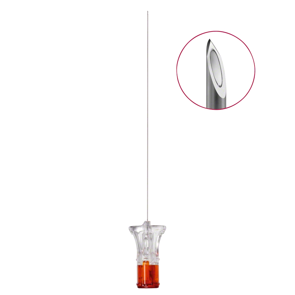
- Spinocan spinal needle
- Quincke type
- Pitkin
Non-cutting Needles with Distal Side Hole “Pencil-Point Spinal Needle”

- Whitacre
- Sprotte
- Gertie Marx
Needle Size: 25-26-27 G (gauge).
Pencil Point spinal needles use an introducer while sharp needles do not. The use of an introducer generally requires initial skin anesthesia with a thin injection needle before the introducer is inserted. The Pencil Point needle is always used for emergency cesarean sections in spinal anesthesia.
Contraindications
- Severely deranged coagulation
- The patient absolutely does not want spinal anesthesia
- Skin infection at the injection site
- Previous EA or spinal complications.
- Neurological deficit symptoms of peripheral origin.
Relative Contraindications
- Neurological dysfunction (MS, myasthenia gravis – however, it is controversial whether a contraindication really exists)
- Spinal tumor, spinal stenosis, or recent trauma to the back
- Sepsis
- Patients with altered consciousness
- Tattoo at the injection site
Coagulation Tests
- Spinal anesthesia causes fewer puncture complications than epidural anesthesia.
- APTT should be normal
- INR > 1.4 and platelet count (PLT) > 100, if comfort gain
- INR > 1.6 or PLT 50-100, if morbidity gain
- INR > 1.8, PLT 30-50, doubtful with spinal anesthesia, if mortality gain.
- If INR > 1.4 or PLT < 100, adjust coagulation before the puncture.
Puncture Level
- Orthopedic extremity surgery, L2-L3, L3-L4, L4-L5.
- Cesarean section L2-L3, L3-L4.
- Peripheral vascular surgery L2-L3, L3-L4.
Landmarks
- Crista iliaca L4


Sensory Assessment – Distribution
- Th 4 – nipple line
- Th 8 – costal margin
- Th 10 – umbilical level
- Th 12 – groin

Bromage Scale

- 0 = can lift the leg with the knee extended
- 1 = can bend at the knee joint
- 2 = can bend at the ankle joint
- 3 = cannot bend at the ankle joint, paralysis
Monitoring
- VAS scale
- Blood pressure and pulse, leg mobility every 4 hours
- Sedation level and respiratory rate once per hour for the first 6 (fentanyl, sufentanil) or 12 hours (morphine) at start-up. When changing dose/bolus, every 2 or 4 hours.
- Then checks every four hours.
Removal of Spinal Catheter
- Usually does not require special sedation.
- Clean the puncture site with alcohol.
- Carefully remove the catheter
- Check the catheter tip after removal
- Keep the urinary catheter for 6 hours
- Keep the IV line for 6 hours
- Continue monitoring for 4 hours
- Wait at least 2 hours before re-administering anticoagulation.
Spinal and Anticoagulation
- LMWH > 5000 E/40 mg – at least 24 hours before spinal anesthesia
- LMWH 2500-5000 – given no later than 12 hours before puncture/manipulation
- IV heparin – wait 3 hours + new APTT
- Single therapy low-dose ASA and/or NSAIDs – if comfort gain
- High-dose ASA – accepted if morbidity gain
- ASA/Plavix – accepted if mortality gain
- Plavix (clopidogrel) should be discontinued at least 5 days before
Failed spinal anesthesia
Spinal doses of anesthetics in different interventions
Spinal doses of anesthetics
| Operation | Bupivacaine Spinal with glucose ("heavy") 5 mg/ml | Bupivacaine Spinal 5 mg/ml | Fentanyl 0,05 mg/ml | Morphine Special 0,4 mg/ml | Other drugs |
|---|---|---|---|---|---|
Gynecology/Obstetrics |
|||||
| Caesarian Sectio | 2,0-2,4 ml | 0,1 ml | 0,25 ml * | ||
| Childbirth injuries suturing | 1 | ||||
| Sphincter rupture | 1,0-1,6 ml | ev. 0,2 ml | |||
| Placental dissolution | 1,2 ml | 0,2 ml | |||
| Childbirth spinal (if parturition is expected < 3 h) | 0,5 ml (alt. 1 ml Bupivacaine 2,5 mg/ml) | Sufentanil 1 ml 5 µg/ml | |||
| Prolaps uteri | 1,8-2,5 ml | 0,2 ml | |||
| Epidural refill for sectio/sphincter rupture (epidural!) | 1 ml (epidural) | Ropivacaine 10-20 ml 7,5 mg/ml | |||
| Vaginal hysterectomy (without GA) | 2,6-2,8 ml | 0,2 ml | 0,25 ml | ||
| Abdominell hysterektomi (med GA) | 2,0-2,5 ml | 0,2 ml | 0,25 ml | ||
|
|||||
| Hip fracture | 1,6-2,2 ml | 0,2-0,4 ml | |||
| Hip joint surgery (possibly CSE) | 1,6-2,2 ml | 0,2-0,4 ml | clonidine 0,4 ml 150 µg/ml if not CSE | ||
| Knee plastic unilateral | 2,6-3,0 ml | 1,6-3,0 ml | |||
| Knee replacement surgery total / revision (possibly CSE) | 1,6-2,4 ml | 0,2-0,4 ml | clonidine 0,4 ml 150 µg/ml if not CSE | ||
| Operation of lower leg, ankle, or foot | 1,8-2,4 ml | 1,8-2,4 ml | ev. 0,2-0,4 ml | alternativt Bupivacaine Spinal | |
| Amputation leg (CSE preferred) | 1,6-2,2 ml | 0,4 ml | ev 0,25 ml if not CSE | ||
| Arthroscopy knee | 1,8-2,4 ml | 1,8-2,4 ml | |||
Urology |
|||||
| TUR-P, TURIS-P | 1,8-2,5 ml | ||||
| TUR-B, Cysto-PX, TUIP, internal urethrotomy | 1,8-2,5 ml | ||||
| TUR-B low dose on day care surgery (1 h block) | 1 ml | Sufentanil 1 ml 5 µg/ml | |||
| Lithotripsy | |||||
| Urethroscopy/Lithotripsy | 2,5-3,0 ml | ev. 0,2 ml | |||
General surgery |
|||||
| Laparoscopic / robot colon surgery Rectum amputation | 1 ml | 0,5 ml | |||
| Inguinal hernia/vascular surgery (operation time < 3 h) | 2,8 ml | ||||
| Peripheral vascular bypass | 3,0-3,2 ml | ev. 0,2 ml | |||
| Inguinal hernia | 2,6-3 ml | ||||
| * not on-call time if the post operation unit cannot monitor for 12 hours | |||||
Spinal Anesthesia – Medications and Dosage
Spinal anesthesia for surgical procedures including orthopedics
| Local anesthetic agent | Drug | Concentration | Dose (ml) | Amount (mg) | Onset Time | Duration |
|---|---|---|---|---|---|---|
Interventions in the lower extremities including hip surgery |
||||||
| Bupivacaine | Marcaine spinal | 5 mg/ml | 2–4 ml | 10–20 mg | 5–15 min | 2–4 hours |
| Bupivacaine | Marcaine spinal Heavy | 5 mg/ml | 2–4 ml | 10–20 mg | 3–15 min | 1,5–3 hours |
| Ropivacaine | Naropine | 5 mg/ml | 3–4 ml | 15–20 mg | 1–5 min | 2–6 hours |
| Levobupivacaine | Chirocaine | 5 mg/ml | 3 ml | 15 mg | ||
Urological surgery |
||||||
| Bupivacaine | Bupivacaine Spinal with glucose ("heavy") 5 mg/ml | 5 mg/ml | 1,5–3 ml | 7,5–15 mg | 5–8 min | 1,5–3 hours |
Abdominal surgery |
||||||
| Bupivacaine Spinal with glucose ("heavy") 5 mg/ml | 5 mg/ml | 2–4 ml | 10–20 mg | 5–8 min | 45–60 min | |
Spinal Anesthesia for Cesarean Sectio
| Drug (Brand name) | Local anesthetics | Concentration | Dose (ml) | Opioid |
|---|---|---|---|---|
| Marcaine Spinal Heavy | Bupivacaine with Glucose | 5 mg/ml | 1,8–2,4 ml (7,5–12,5 mg) | |
| Marcaine Spinal Heavy | Bupivacaine with Glucose | 5 mg/ml | 1,8-2,4 ml + | Fentanyl 15-25 μg |
| Marcaine Spinal Heavy | Bupivacaine with Glucose | 5 mg/ml | 1,8-2,4 ml + | Morphine 0,1 mg (0.4 mg/ml 0.25 ml) |
| Marcaine Spinal Heavy | Bupivacaine with Glucose | 5 mg/ml | 1,8-2,4 ml + | Fentanyl 15-25 μgram + Morphine 0,1 mg (0,4 mg/ml 0,25 ml) |
| Naropin | Ropivacaine | 5 mg/ml | 1,5-3 ml (7,5-15 mg) |
Addition of opioids in spinal anesthesia
| Local anesthetic | Concentration | Dose (weight units) | Dose in ml |
|---|---|---|---|
| Morpine | 0.4 mg/ml | 0,1–0,2 mg | 0,25–0,5 ml |
| Fentanyl | 50 mikorg/ml | 20–40 μg | 0,4–0,8 ml |
| Sufentanil | 5 μg/ml | 2,5–5–10 μg | 1–1,5 ml |
Spinal Anesthesia for Labor Pain
| Brand Name | Local anesthetic | Concentration | Dose (ml) | Opioid |
|---|---|---|---|---|
| Naropin | Ropivacaine | 5 mg/ml | 0,2-0,3 ml (1-1,25 mg) | Sufentanil 5 μg/ml 1-1,5 ml (7,5 mikrog) |
| Marcain Spinal | Bupivacaine | 5 mg/ml | 0,2-0,4 ml (1-2 mg) | Sufentanil 5 μg/ml 1-1,5 ml (7,5 mikrog) |
Bupivacaine (Marcaine) Spinal 5 mg/ml
- Procedures in the lower extremities including hip surgery.
- Dosage: 2-4 ml, 10-20 mg.
- Onset time 5-8 minutes.
- Duration 1.5-4 hours. Recommended injection site is below L3.
Bupivacaine (Marcaine) Spinal Heavy 5 mg/ml
- Procedures in the lower extremities including hip surgery.
- Dosage: 2-4 ml, 10-20 mg. Onset time 5-8 minutes. Duration 1.5-4 hours.
- Urological surgery: Dosage: 1.5-3 ml, 7.5-15 mg. Onset time 5-8 minutes. Duration 2-3 hours. Recommended injection site is below L3.
- Abdominal surgery: Dosage: 2-4 ml, 10-20 mg. Onset time 5-8 minutes. Duration 45-60 minutes.
Ropivacaine (Naropin) 5 mg/ml
- Procedures in the lower extremities including hip surgery.
- Dosage: 3-4 ml, 15-20 mg.
- Onset time 1-5 minutes.
- Duration 2-6 hours.
Levobupivacaine (Chirocaine) 5 mg/ml
- Procedures in the lower extremities including hip surgery.
- Dosage: 3 ml, 15 mg.
Cheat Sheet Spinal Medication Doses [Swe]

Click on the image to download the PDF file
Spinal anesthesia in morbid obesitas
Postoperative Monitoring After Spinal Anesthesia
Expected (but not guaranteed) motor block after spinal anesthesia:
- Bupivacaine (Marcaine) Heavy: 2 hours
- Bupivacaine (Marcaine) Spinal: 3 hours
- Bupivacaine (Marcaine) Spinal + Clonidin (Catapres): approx. 5 hours.
Monitoring times take into account the following additives of spinally administered medications
Recommended monitoring times after spinal anesthesia
| Respiratory monitoring | Monitoring of motor skills according to Bromage | Bromage-scale | |
|---|---|---|---|
| Bupivacaine | - | 8 hours | 0 = can lift the leg with the knee extended |
| Fentanyl | 6 hours | - | 1 = can bend at the knee joint |
| Morphine | 12 hours | - | 2 = can bend at ankle |
| Clonidine | - | 10 hours | 3 = cannot bend the ankle, paralysis |
Knudsen’s Test in accidental dura punction
Knudsen’s test is a simple bedside examination that can detect the presence of cerebrospinal fluid. It can be used when spinal puncture of an EPi catheter or skull base fracture with cerebrospinal fluid leakage is suspected. One takes a cotton swab or a compress and gently rubs it with colored chlorhexidine solution. Then, the fluid to be examined is applied to the swab. If the color changes to red, cerebrospinal fluid is present in the sample, indicating a positive Knudsen’s test. The last image shows the result of an examination with saliva from a patient with a skull base fracture and cerebrospinal fluid leakage. The comparison swab is stained with normal saliva.


- Knudsen K. Coloured chlorhexidine cotton pads may help to identify cerebrospinal fluid during epidural or spinal anaesthesia. Acta Anaesthesiol Scand. 2006 Jul;50(6):685-7. doi: 10.1111/j.1399-6576.2006.01046.x. PMID: 16987362.
Intrathecal Pain Relief
Intrathecal pain treatment is administered by inserting an epidural catheter under strict sterile conditions into the spinal space via a lumbar puncture with an epidural needle. The catheter is then used for continuous spinal anesthesia via a pump – e.g., Gemstarpump or CAD-pump. This puncture is performed like a spinal anesthesia with an epidural needle. Through the epidural needle, the spinal space is first identified with a spinal needle, i.e., a “needle-through-needle technique”. The epidural catheter is then inserted high up, about 15 cm after the insertion in L2-L3. The catheter can advantageously be tunneled subcutaneously from the insertion site to the side of the patient. Intrathecally administered medication is 100–300 times stronger than orally administered.

Contraindications
High intracranial pressure, sepsis, severe coagulation disorder, increased risk of bleeding, anticoagulant treatment (Warfarin, Plavix), and infected skin, e.g., pressure sores near the insertion area.
Preoperative Blood Tests
INR, APTT, platelets, CRP.
Anticoagulated Patients
Warfarin treatment is discontinued – the operating physician informs of the acceptable INR in each individual case. Patients treated with low molecular weight heparin, e.g., Fragmin, should be treated as follows: With Fragmin 5000 E s.c. injection, 10 hours should pass before insertion, position adjustment, or removal. The subsequent dose of Fragmin is given 2 hours after insertion or removal of the intrathecal catheter.
With Clexane 40 mg s.c., 10 hours should pass before insertion, position adjustment, or removal. The subsequent dose of Clexane is given 2 hours after insertion, position adjustment, or removal of the intrathecal catheter. Plavix should be discontinued 10 days before the procedure.
Medications for Intrathecal Administration
95 ml Bupivacaine (Marcaine) 5 mg/ml is mixed with 5 ml Morphine 10 mg/ml to a volume of 100 ml. The concentration of Bupivacaine becomes 4.75 mg/ml and the concentration of morphine becomes 0.5 mg/ml. A recommended starting dose is 0.3 ml/hour with the option to give a bolus dose of 0.2 ml if needed.
Perioperative pain relief is usually achieved by administering local anesthetics with the addition of opioids, usually morphine 0.1 mg, 100 μg.
There is the option to administer a mixture of opioids and clonidine (Catapresan). Such a mixture can be, for example:
- Morphine Special 0.1-0.3 mg, 0.4 mg/ml, 0.25–0.75 ml
- Sufentanil (Sufenta) 10 μg, 5 μg/ml, 2 ml
- Clonidin (Catapres) 75 μg, 150 μg/ml, 0.5 ml
Monitoring for Intrathecal (Subarachnoid) Administration of Opioid
After morphine is given intrathecally, the following checks are performed for 12 hours, and after intrathecal fentanyl or sufentanil is given for 6 hours.
One time per hour after spinal anesthesia is given, the following are checked:
- VAS
- Sedation level
- Respiratory rate (if sedation level >1)
- Nausea
- Itching
Subsequent checks according to the above are performed every 4 hours for an additional 12 hours.
Extra check twice per hour for 2 hours when adding sedative or respiratory depressant medications.
Urinary catheter for at least 12 hours. Check bladder function after the urinary catheter is removed.
PDPH (Post Dural Puncture Headache)
Definition
PDPH is defined according to international classification as a posture-dependent headache that occurs within 5 days after a dural puncture and resolves spontaneously within a week or within 48 hours after an epidural blood patch. It is also associated with neck stiffness, tinnitus, hearing changes, light sensitivity, and nausea (1).
Background
After puncture of the dura mater, the tough outer membrane of the brain, a cerebrospinal fluid leak into the epidural space may occur. This can cause intracranial hypovolemia, with traction on cranial structures. A headache may occur, but the exact mechanism of the symptoms is not known. The intracranial hypovolemia can lead to cranial nerve impairment and venous dilation, thus posing a risk of developing a subdural hematoma.
The severity of the headache is influenced by the needle’s size and design, the patient’s age and gender, with the greatest risk in young women after puncture with a large needle. Obstetric patients are therefore a risk group considering age, gender, and the frequency of epidural anesthesia during childbirth. The risk of PDPH after puncture with a 27 G pencil point spinal needle is estimated at between 0-2%, while the risk of PDPH after accidental puncture with a 16-18 G epidural needle is 45-80% (2, 3). The incidence of accidental dural puncture during labor epidural anesthesia is about 1% (4).
Symptoms
The typical symptoms consist of severe posture-dependent headache. It occurs in 90% of cases within 3 days after the puncture (2, 5). The headache is usually frontal but can also be located in the neck with radiation to the shoulders. The headache worsens in an upright position and is significantly relieved when lying down. Nausea, dizziness, tinnitus, hearing impairment, and light sensitivity are relatively common symptoms (2, 4).
Differential Diagnosis
Intracranial mass effect (e.g., bleeding, tumor), cerebral venous thrombosis, migraine, infectious meningitis, tension headache, preeclampsia, anemia, Sheehan’s syndrome, Posterior Reversible Encephalopathy Syndrome (PRES) (2, 3, 6). Prognosis In most cases, the symptoms resolve spontaneously within a week (2). However, chronic symptoms can also occur. Complications in the form of cranial nerve impairment and subdural hematoma can occur (6, 7). In one case series, the incidence of subdural hematoma was 0.026% in an unselected obstetric epidural anesthetized population and 1.1% (1 in 87) in the group where dural puncture was confirmed in connection with the administration of spinal anesthesia (8).
Treatment
Many different treatments have been tried to relieve this intense headache. Professional support, careful explanation of the symptoms, and follow-up are important. Although the symptoms are relieved in a lying position, bed rest has not been shown to affect the duration (2). Avoiding dehydration, administering analgesics, triptans, antiemetics, and acupuncture can relieve symptoms but rarely provide complete symptom relief. These methods can be tried in mild cases and/or if there is a contraindication for treatment with a blood patch. Some effect has been shown with prophylactically administered cosyntropin IV and epidurally administered morphine (9, 10). The most effective treatment to date is an epidural blood patch.
Treatment of PDPH (Post Dural Puncture Headache)
Treatment of PDPH with Epidural Blood Patch
The method involves an anesthesiologist injecting 20 ml of autologous blood into the epidural space at the same level as the dural puncture or one interstitium below. Theoretically, a momentary increase in volume and pressure in the spinal canal is achieved with subsequent reduction of traction on intracranial structures. This leads to a rapid reduction in headache. The blood then spreads relatively quickly in the epidural space, especially in the cranial direction. The blood forms a “clot” at the tissue injury in the dura and seals the hole (2, 3, 11). The epidural blood patch was first described in the 1960s, and much of the knowledge about the treatment is based on case reports and retrospective studies. Diverging recommendations exist regarding the volume of blood injected, the timing of treatment, and whether prophylactic/therapeutic treatment should be applied (2, 3, 12, 13). There is some support that therapeutic treatment should be administered no earlier than 24 hours after the puncture to reduce the need for repeated treatment (3, 14).
Indication
Debilitating, classic, posture-dependent headache after spinal anesthesia, where other differential diagnoses have been considered but excluded. Investigation: Blood pressure, temperature, hemoglobin, white blood cell count, and CRP.
Contraindication
As usual for spinal anesthesia: coagulation disorder, anticoagulant treatment, sepsis, infection at the puncture site, patient refusal. In the case of bloodborne infections in the patient, individual assessment must be made.
Method
Under sterile conditions, the epidural space is located using the usual epidural needle and technique, preferably one interstitium caudal to the previous puncture. The assistant (usually a nurse) then performs a sterile venipuncture and aspirates at least 20 ml of autologous blood from the patient. The blood syringe is handed directly to the anesthesiologist for immediate slow injection of blood (up to 20 ml) into the epidural space. The injection should be stopped if the patient reports back pain. Apply a dressing to the puncture site. Bed rest is recommended for 1-2 hours, followed by cautious mobilization.
Relief from headache is achieved in >70% of PDPH patients treated with a blood patch (2, 3). If the headache recurs with typical symptoms, the blood patch treatment may need to be repeated (4, 5).
As the diagnosis of PDPH is primarily clinical, differential diagnoses must be considered on broad indications. In cases of atypical, persistent, or recurrent symptoms, altered type of headache, and/or the development of other neurological symptoms, further investigation with e.g., CT/MRI must be carried out (2, 6, 8).
Risks/Complications
There is a risk of further dural puncture in connection with the localization of the epidural space. Mild back pain lasting up to 5 days occurs (3). Infection in the spinal canal. If the blood is accidentally injected subdurally/intradurally, it can cause serious complications in the form of arachnoiditis, cauda equina syndrome, and permanent nerve damage (3, 15, 16).
After PDPH and/or a blood patch, the patient must be closely monitored and must not leave the hospital if severe headache persists. Detailed information, both verbal and written, regarding the complication and its risks, must be provided. Clear instructions on where the patient should turn in case of recurrent symptoms. The complication must be documented in the medical record.
In 2016, a European observational study, EPiMAP, was initiated. This will hopefully provide us with more knowledge about blood patches as a treatment for PDPH.
References:
- International classification of headache disorders 2nd edition. Cephalgia: 2004, 24: 79.
- Post-dural puncture headache: pathogenesis, prevention and treatment. BJA (2003) 91 (5):718-729.
- Post-dural puncture headache: The worst common complication in obstetric anesthesia. Seminars in perinatology: vol 38 Issue 6 (2014) 386-394.
- Management of accidental dural puncture and post-dural puncture headache after labor: a Nordic survey. Acta Anaesth Scand 2011; 55: 46–53.
- Ten years of experience with accidental dural puncture and post-dural puncture headache in a tertiary obstetric anesthesia department. IJOA: (2009) 17; 329-335.
- Intracranial subdural hematoma following neuraxial anesthesia in the obstetric population: a literature review with analysis of 56 reported cases. IJOA vol 25 Feb 2016, 58-65.
- MBRRACE report 2009-2011 (2012).
- Subdural Hematoma Associated With Labor Epidural Analgesia: A Case Series. Reg Anesth and Pain Medicine: 2016: vol 41(5) 628-631.
- Prevention of post-dural puncture headache in parturients: a systematic review and meta-analysis. Acta Anaesth Scand 2013:57:417-430.
- Cosyntropin for prophylaxis against Post Dural Puncture Headache after Accidental Dural Puncture. Anesthesiology: 2010, vol 113(2)413-420.
- Magnetic resonance imaging of extradural blood patches: appearances from 30 min to 18h. BJA: 1993:71(2) 182-188.
- Prophylactic vs therapeutic blood patch for obstetric patients with accidental dural puncture-a randomized controlled trial. Anesthesia 2014, 69, 320-326.
- Accidental dural puncture, postdural puncture headache, intrathecal catheters and epidural blood-patch: revisiting the old nemesis. J Anesth (2014) 28:628-630.
- The influence of timing on the effectiveness of epidural blood patches in parturients. IJOA (2013) 22, 303-309.
- Spinal subdural hematoma after an epidural blood patch. IJOA: vol 24 Issue 3 Aug 2015, 288-289.
- Chronic adhesive arachnoiditis after repeat epidural blood patch. IJOA: vol 24 Issue 3 Aug 2015, 280-283.
Disclaimer:
The content on AnesthGuide.com is intended for use by medical professionals and is based on practices and guidelines within the Swedish healthcare context.
While all articles are reviewed by experienced professionals, the information provided may not be error-free or universally applicable.
Users are advised to always apply their professional judgment and consult relevant local guidelines.
By using this site, you agree to our Terms of Use.
